Venous Disease
Thrombophlebitis, chronic venous insufficiency, varicose veins, and post-thrombotic syndrome
Clinical Evidence — Not FDA-Evaluated
Historical clinical practice describes benefit from hirudotherapy in superficial thrombophlebitis, chronic venous insufficiency, and varicose ulcers, but these reports are non-PubMed-indexed and not independently verifiable, and no PubMed-indexed controlled trial establishes efficacy in these indications. The strongest venous-disease-specific study is a single small, uncontrolled prospective series (Bapat et al., 1998, PMID 9701897), whose authors themselves call for controlled trials. These specific applications are not FDA-cleared, though the venous decompression mechanism directly underlies the FDA 510(k)-cleared microsurgical indication.
Investigational Application
International Clinical Evidence
The strongest venous-disease-specific study is a single small, uncontrolled prospective series in complicated varicose veins (Bapat et al., 1998, n=20), whose authors themselves call for controlled trials. Objective microcirculation data come from a small instrumental study in congested tissue (Rothenberger et al., 2016, n=12), and the largest synthesis (Whitaker et al., 2012) concerns venous congestion in microsurgical flaps rather than venous disease. Standard vein care — compression therapy and, where indicated, definitive vascular intervention — remains primary; hirudotherapy for venous disease is investigational.
Biological Mechanisms: SGS Effects on the Venous System
The therapeutic effect of hirudotherapy in venous disease operates through multiple simultaneous pathways that no single pharmaceutical agent replicates. These mechanisms have been characterized through laboratory studies of individual salivary gland secretion (SGS) components, instrumental monitoring of microcirculation and tissue oxygenation, and serial hemostatic parameter measurements in clinical populations.
Venous Decompression
- Blood extraction: Each leech ingests 5-15 mL during a 30-90 minute feeding, creating immediate mechanical decompression of congested venous tissue
- Sustained post-detachment bleeding: The bite wound continues to ooze 10-50 mL over 4–24 hours after leech detachment, mediated by persistent hirudin anticoagulation and calin-mediated platelet inhibition
- Net volume reduction: 15-65 mL total blood removal per leech application (feeding + post-detachment oozing), directly reducing venous pressure in the treated vascular bed
- Targeted extraction: Bapat et al. (1998) documented that leech-extracted blood pO2 (40.05 mm Hg) exceeds venous blood pO2 (34.33 mm Hg), confirming preferential extraction from the congested venous compartment
Microcirculation Evidence
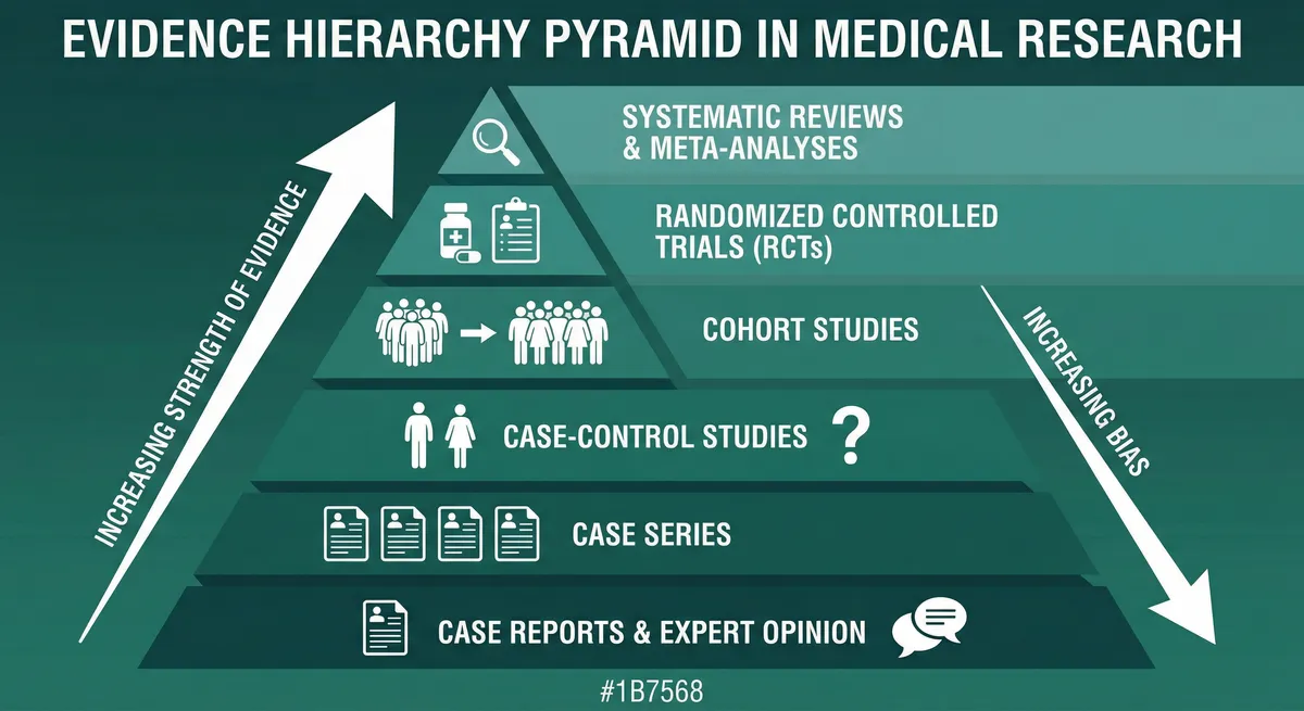
GRADE Evidence Level: Moderate
RCTs with limitations or strong observational studies
The microcirculatory effects of hirudotherapy have been documented instrumentally through tissue spectrophotometry, laser Doppler flowmetry, and partial oxygen pressure (pO2) measurement of leech-extracted blood. These objective measurements confirm that leech application produces measurable, if transient, improvements in local blood flow and tissue oxygenation — the fundamental mechanisms underlying any benefit in venous disease.
Rothenberger et al. (2016) applied a single leech to venous-congested tissue in 12 patients and measured perfusion with an Oxygen-to-See (O2C) tissue spectrophotometry and laser Doppler device. One hour after application, blood flow rose 56.7%, relative hemoglobin fell 25.5%, and tissue oxygen saturation rose 53.7% (all statistically significant); the values returned to baseline by three hours.
Of particular relevance to venous disease is the pO2 measurement in the study by Bapat et al. (1998). Comparing oxygen tension in arterial blood, venous blood, and blood sucked by leeches in 7 patients, the investigators found the mean pO2 of leech-sucked blood (40.05 +/- 7.24 mm Hg) was similar to that of venous blood (34.33 +/- 8.40 mm Hg). On this basis the authors concluded that the medicinal leech sucks venous blood, consistent with local venous decompression.
| Study | Design | Population (n=) | Intervention | Key Outcome | Result |
|---|---|---|---|---|---|
| Rothenberger et al. 2016 | Prospective instrumental study | Patients with venous-congested tissue requiring leech therapy (n=12) | Leech application with perfusion measured by Oxygen-to-See (O2C) tissue spectrophotometry and laser Doppler flowmetry before and at 10 min, 1 h, and 3 h | Blood flow, relative hemoglobin, and tissue oxygen saturation over time | One hour after leech application: blood flow +56.7%, relative hemoglobin -25.5%, oxygen saturation +53.7% (all significant). Values returned to baseline by 3 hours Objective instrumental confirmation that a single leech relieves venous congestion for roughly one hour. Indexed in PubMed (PMID 27733014) |
| Bapat et al. 1998 | pO2 substudy (within n=20 series) | Complicated varicose vein patients (subset of 7) (n=7) | pO2 measured in arterial blood, venous blood, and blood sucked by leeches | Comparison of oxygen tension across blood compartments | Mean pO2 of leech-sucked blood was 40.05 +/- 7.24 mm Hg, similar to venous blood (34.33 +/- 8.4 mm Hg) The authors conclude the medicinal leech sucks venous blood. No arterial-comparison intermediate value or capillary-admixture claim is supported by the paper. Indexed in PubMed (PMID 9701897) |
Targeted Venous Decompression
Rheological Effects: Blood Viscosity, Fibrinogen, and Platelet Function
GRADE Evidence Level: Moderate
RCTs with limitations or strong observational studies
Clinical evidence suggests that hirudotherapy produces measurable improvements in blood rheological parameters that are directly relevant to venous disease pathophysiology. Elevated blood viscosity, increased fibrinogen levels, and enhanced platelet aggregation are established risk factors for venous thrombosis and contribute to the progression of chronic venous insufficiency through impaired microcirculatory flow. Peer-reviewed studies report substantial and sustained changes in these parameters following hirudotherapy.
Quantitative Rheological Changes
- Fibrinogen reduction: reductions in fibrinogen have been described in clinical practice, but the specific magnitudes and durations reported in the older literature are not independently verifiable and are not established by any PubMed-indexed controlled study
- Whole blood viscosity: reported to decrease below baseline in the days following treatment and to recover over subsequent weeks in clinical accounts; these figures are not independently verifiable, suggesting repeated sessions if a sustained effect is sought
- Platelet aggregation: reported to decrease after treatment in clinical accounts, consistent with the combined antiplatelet action of calin (collagen-mediated pathway) and other SGS compounds; the specific figures are not independently verifiable
- Lymphogram coagulation time: prolongation of coagulation parameters after leech therapy has been described in clinical practice; the specific values reported are not independently verifiable
Post-Thrombotic Syndrome
GRADE Evidence Level: Low
Observational studies or RCTs with serious limitations
Varicose vein treatment with medicinal leeches has historical roots predating modern vascular surgery and remains part of international clinical practice. Approximately 10-17% of the global adult population is affected by varicose vein disease, with complications that include superficial thrombophlebitis, lymphangitis, chronic venous ulceration, and progression to post-thrombotic syndrome. Positive symptomatic responses have been reported in clinical practice for uncomplicated peripheral venous disease, but these older reports are non-PubMed-indexed and not independently verifiable.
Clinical experience suggests symptomatic improvement — reduced heaviness, aching, edema, and skin changes — following leech application to the varicose vein course. The proposed mechanism involves direct venous pressure reduction through blood extraction and sustained post-detachment bleeding, combined with the anti-inflammatory and rheological effects of SGS. Application sites include the course of the affected vein, the ankle region, and areas of maximal edema.
Post-Thrombotic Syndrome: Efficacy Unproven
- No verifiable clinical series: there is no PubMed-indexed clinical trial or case series establishing leech therapy efficacy for post-thrombotic syndrome
- Mechanistic rationale only: any rationale rests on the general venous-decompression mechanism, not on direct outcome data in PTS
- Standard care remains primary: compression therapy and management of the underlying venous disease are the established approach to PTS
- Research gap: a prospective controlled study using the Villalta scale would be required before any clinical recommendation
Deep Vein Thrombosis: Important Limitations
Deep vein thrombosis of the lower extremities is a potentially life-threatening condition, with pulmonary thromboembolism representing the most serious complication and a 30-day case fatality rate of approximately 6% (Kahn et al., 2014). While the pharmacologic rationale for hirudotherapy in DVT is sound — the combination of hirudin (thrombin inhibition), destabilase (fibrinolysis), and calin (antiplatelet activity) addresses all three arms of Virchow's triad — critical limitations must be explicitly stated.
DVT: Not a Substitute for Standard Anticoagulation
What Hirudotherapy Does NOT Replace in DVT
- Duplex ultrasound diagnosis and monitoring
- LMWH or DOAC anticoagulation per current guidelines (ACCP, ASH, ESC)
- Risk stratification for pulmonary embolism (Wells score, Geneva score)
- Catheter-directed thrombolysis in selected cases
- IVC filter placement when anticoagulation is contraindicated
- Compression therapy for post-thrombotic prevention
Potential Adjunctive Rationale (Theoretical)
- Multi-target antithrombotic action addressing stasis, endothelial injury, and hypercoagulability simultaneously
- Destabilase-mediated thrombolysis via a unique isopeptidase mechanism distinct from plasmin
- Anti-inflammatory activity reducing venous wall damage and potentially lowering PTS risk
- No clinical trial data to support these theoretical benefits in DVT specifically
Safety and Drug Interactions for Venous Disease Patients
Venous disease patients present specific safety considerations for hirudotherapy, primarily related to the high prevalence of concurrent anticoagulant and antiplatelet therapy in this population. The additive anticoagulant effect of SGS components — particularly hirudin (direct thrombin inhibitor) and calin (platelet aggregation inhibitor) — combined with systemic anticoagulation creates an elevated bleeding risk that requires careful management, enhanced monitoring, and documented informed consent.
Anticoagulant Interaction Warning
| Drug | Class | Interaction | Management |
|---|---|---|---|
| Warfarin | Vitamin K antagonist | Additive anticoagulant effect with hirudin; prolonged and excessive bleeding from bite sites | Check INR before application; target INR at lower end of therapeutic range if possible; enhanced bleeding monitoring; low transfusion threshold |
| Apixaban, rivaroxaban, edoxaban | DOACs (Factor Xa inhibitors) | Additive anticoagulation; no readily available reversal agent at many facilities | Consider timing relative to DOAC trough; andexanet alfa for life-threatening bleeding; enhanced monitoring |
| Dabigatran | DOAC (direct thrombin inhibitor) | Mechanistically redundant with hirudin (both inhibit thrombin); highest theoretical bleeding risk among DOACs | Idarucizumab available for reversal; consider holding dose if clinically feasible; closest monitoring required |
| Heparin (UFH) | Indirect thrombin inhibitor | Additive anticoagulation; commonly administered concurrently in microsurgical settings (54.29% of patients per Whitaker 2012) | Titrate to aPTT; institutional protocol for concurrent use; serial hematocrits |
| Enoxaparin, dalteparin | LMWH (Factor Xa via AT) | Additive anticoagulation; predictable pharmacokinetics | Standard dosing generally acceptable with monitoring; avoid within 12 hours of leech application if bleeding is excessive |
| Aspirin | Irreversible COX-1 inhibitor | Additive antiplatelet effect with calin; prolonged bleeding time | Do not discontinue in patients with cardiovascular indications; enhanced bleeding monitoring |
| Clopidogrel, prasugrel, ticagrelor | P2Y12 receptor antagonists | Additive antiplatelet effect with calin; enhanced bleeding risk | Enhanced monitoring; involve cardiology in risk-benefit discussion before leech therapy |
| Study | Design | Population (n=) | Intervention | Key Outcome | Result |
|---|---|---|---|---|---|
| Mumcuoglu et al. 2014 | Review of contraindications | Patients receiving medicinal leech therapy (n=NR) | Review of recommendations, contraindications, and adverse events of leech therapy | Contraindications and safety considerations | Describes contraindications including bleeding disorders, severe anemia, and immunosuppression, and highlights bleeding and infection (Aeromonas) risks General leech-therapy safety reference, not venous-disease-specific |
| Whitaker et al. 2012 | Systematic review | Plastic/reconstructive surgery patients receiving leech therapy for venous congestion (n=277 cases (67 papers)) | Medicinal leech therapy for venous congestion, with concurrent anticoagulation in many cases | Success (flap/tissue salvage), transfusion, antibiotics, and concurrent anticoagulation rates | Overall success 77.98% (216/277); 49.75% required transfusion; 79.05% received antibiotics; 54.29% received concurrent anticoagulant therapy; overall complication rate 21.8% Largest synthesis of leech therapy for venous congestion, but in microsurgery, not venous disease. Indexed in PubMed (PMID 22407551) |
Absolute Contraindications
- Hemophilia or severe coagulopathy (uncontrollable hemorrhage from bite wounds)
- Hemorrhagic diathesis (including severe von Willebrand disease, factor deficiencies with bleeding phenotype)
- Severe anemia (hemoglobin < 8 g/dL) — insufficient oxygen-carrying capacity to tolerate 15-65 mL additional blood loss per leech
- Documented allergy to leech SGS (risk of anaphylaxis)
- Patient refusal after informed consent discussion
Evidence Gaps and Research Priorities
Venous disease represents a natural target for prospective controlled trials, as standardized endpoints and validated outcome instruments already exist. The direct relationship to the FDA-cleared venous decompression mechanism strengthens the regulatory pathway for investigational studies. However, significant evidence gaps remain in the current literature.
Current Evidence Limitations
- No RCTs for CVI or varicose veins: All CVI data are from uncontrolled or historical, non-PubMed-indexed case reports. No controlled trial establishes efficacy in venous disease; the only prospective venous-disease study is the small uncontrolled Bapat 1998 series
- Small, mostly uncontrolled evidence: the only prospective venous-disease study (Bapat 1998, n=20) is uncontrolled; the largest synthesis (Whitaker 2012, 277 cases) concerns venous congestion in microsurgical flaps, not venous disease
- Absence of validated outcome instruments: Published studies predate modern validated instruments such as VCSS (Venous Clinical Severity Score), CEAP classification, and disease-specific quality of life measures
- Lack of duplex ultrasound confirmation: Objective documentation of thrombus resolution and venous reflux changes using modern imaging is absent from the published literature
- Incompletely characterized rheological data: The rheological figures in the older literature come from non-PubMed-indexed sources that are not independently verifiable; modern hemorheology instruments (rotational viscometry, ektacytometry) would be required to provide reliable measurements
Priority Research Directions
- RCT for superficial thrombophlebitis: Standardized endpoints (resolution time, recurrence rate, quality of life) with duplex ultrasound confirmation of thrombus resolution
- Controlled trial for CVI (CEAP C4-C6): Using VCSS as the primary endpoint, with secondary endpoints of ulcer healing rate, edema reduction, and quality of life (CIVIQ-20)
- Laser Doppler and transcutaneous oximetry: Systematic microcirculation assessment before, during, and after leech application in venous disease patients, with standardized protocols
- Modern rheological profiling: Rotational viscometry, erythrocyte deformability measurement, and complete fibrinolytic panel in a venous disease population
- Combination therapy studies: Hirudotherapy + compression + standard pharmacotherapy versus compression + standard pharmacotherapy alone
- PTS prevention: Prospective study of post-DVT hirudotherapy as adjunctive therapy for reducing PTS incidence, using the Villalta scale
Regulatory Pathway
Key Takeaways
Clinical Evidence Summary
- Historical, non-PubMed-indexed clinical accounts describe faster symptom resolution and shorter recovery for acute thrombophlebitis with hirudotherapy added to standard care, but these reports are not independently verifiable and no PubMed-indexed controlled trial substantiates a specific reduction in hospital stay
- All varicose ulcers healed in a small uncontrolled 20-patient series (95% edema reduction, 75% decreased hyperpigmentation); the authors call for controlled trials (Bapat et al., 1998)
- Post-thrombotic syndrome efficacy is unproven — no PubMed-indexed clinical series supports leech therapy for PTS, and standard compression care remains primary
- Objective instrumental data (Rothenberger et al., 2016, n=12) show local blood flow +56.7% and oxygen saturation +53.7% one hour after a single leech, returning to baseline by 3 hours
- Standard vein care — compression therapy and, where indicated, definitive vascular intervention — remains primary; hirudotherapy for venous disease is investigational
Related Resources
Safety & Infection Control
Complete safety framework including Aeromonas prophylaxis.
All Clinical Specialties
Complete index of 14 medical specialties with evidence tiers.
Coverage Map
Live index of all conditions, compounds, RCTs, and jurisdictions covered on ASH.
How We Grade Evidence
ASH editorial methodology: GRADE framework and claim-boundary taxonomy.